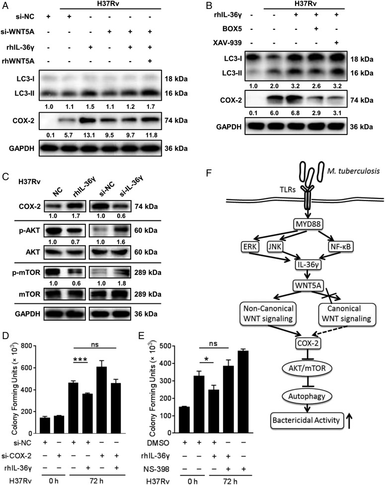
FIGURE 8.
Requirement for WNT5A in IL-36γ mediated autophagic killing of H37Rv. (A and B) LC3-II and COX-2 levels in MDMs transfected with si-WNT5A or a control siRNA before infection with H37Rv and stimulated with rhIL-36γ and/or rhWNT5A (A), or incubated with BOX5 or XAV-939 before infection with H37Rv and stimulated with combination of rhIL-36γ and BOX5/XAV-939 (B), were measured by Western blot. (C) Protein levels of COX-2, p-AKT/AKT, and p-mTOR/mTOR in MDMs infected with H37Rv and treated with rhIL-36γ (left), or transfected with IL-36γ siRNA (right), were measured by Western blot. (D and E) The numbers of viable intracellular bacteria in MDMs infected with H37Rv and transfected with COX-2 siRNA or combined with rhIL-36γ treatment (D), or pretreated with NS-398 with DMSO as the solvent control before infection with H37Rv and then treated with combination of rhIL-36γ and NS-398 (E), were determined using CFU assays. (F) Illustration of model of IL-36γ promotes killing of M. tuberculosis by macrophages via the WNT5A-induced noncanonical WNT signaling pathway. Data presented are from one of least three independent experiments with similar results, and data are shown as means ± SD. *p ≤ 0.05, ***p ≤ 0.001.

