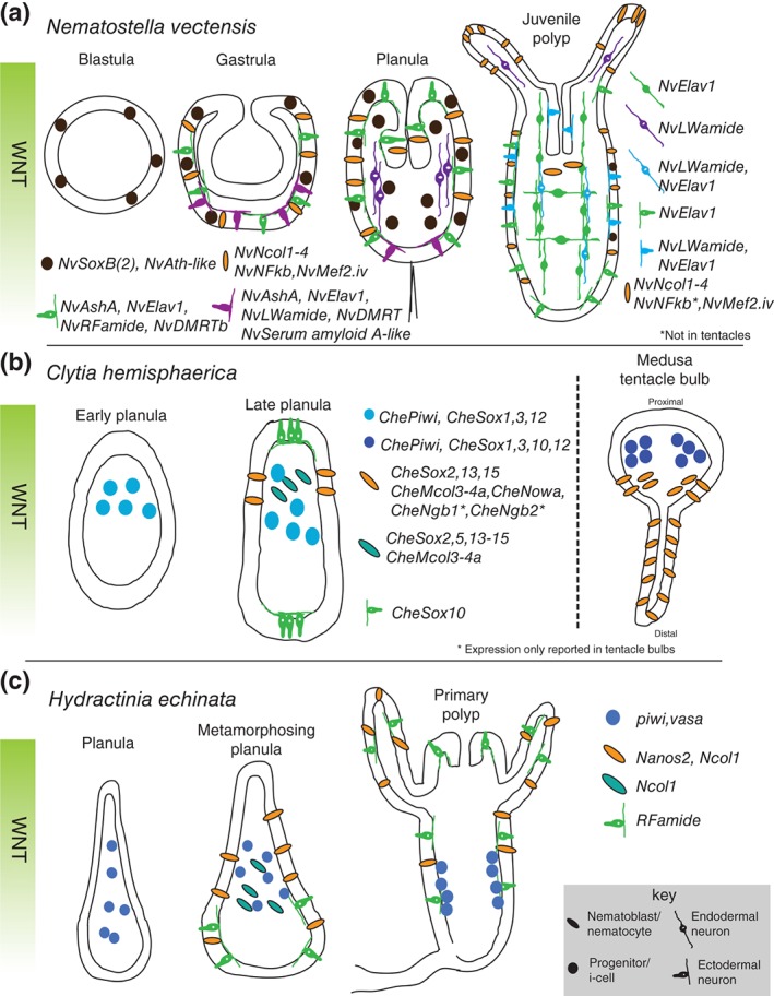
Figure 2.

Summary of neural gene expression during embryogenesis. Some of the known neural gene expression patterns for Nematostella vectensis (a), Clytia hemisphaerica (b), and Hydractinia echinata (c) are shown. Developmental time progresses from left to right with depicted stage indicated above each image. In all planulae and polyp stages, the images are oriented with oral pole facing up. The orientation of the Clytia tentacle (b, right side) is proximal up and distal down. Note that the expression patterns of CheSox genes are depicted in a simplified manner.77 CheNgb, neuroglobin. 78
