
Figure 3.
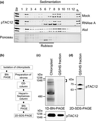
Both ZmpTAC12 isoforms are components of the PEP‐complex. (a) Sucrose‐gradient sedimentation demonstrating that ZmpTAC12 is associated in high molecular weight complexes containing nucleic acids. Stromal extracts of 7‐d‐old maize seedlings were sedimented through sucrose gradients after treatment with RNase A, or AluI, or incubated without nucleases (mock) using identical conditions. Equal proportions of gradient fractions were analyzed by immunoblotting with the indicated antibodies. Ponceau staining is shown to demonstrate the position of Rubisco (c. 550 kDa). Str, stroma; P, pelleted material. (b) Scheme of subcellular fractionation and purification. Intact chloroplasts were isolated from 10‐d‐old seedlings and lysed for stroma preparation. Transcriptionally active fractions were subsequently enriched by QS and HS chromatography, and analyzed by BN/SDS‐PAGE. (c) Blue native gel in the first dimension. Protein complexes from chloroplasts (50 μg crude chloroplast proteins, Cp) and the enriched QS/HS fraction were separated by BN‐PAGE (upper panel) and subsequently immunoblotted with the pTAC12 antibody (lower panel). Immunoblot corresponds to the upper part of the BN gel. Sizes of marker proteins are given on the left. (d) Immunoblot showing both ZmpTAC12 isoforms in PEP enriched fractions. The indicated gel strip (top bracket in (c); c. 1000 kDa) was incubated with denaturing buffer and a 10% SDS‐gel was run in the second dimension (2D‐SDS‐PAGE). ZmpTAC12 isoforms were examined by immunodetection with the anti‐pTAC12 antibody. Numbers on the left represent Mr of molecular weight standards.