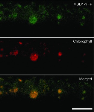
Figure 4.

Expression and localization of MSD1‐YFP in rice cells. Rice cells bombarded with MSD1‐YFP were observed by laser scanning microscopy. Top: MSD1‐YFP; middle: chlorophyll autofluorescence; bottom: merged. Panels are stacks of 30 images per cell, acquired from the top to the middle of the cell, every 1–2 μm. MSD1‐YFP colocalized with chlorophyll autofluorescence. Bar = 10 μm.
