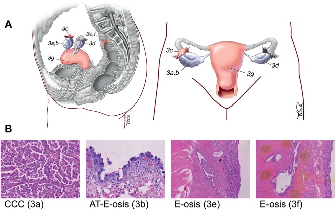
Figure 3.

Specimens collected from case 3. (A) Anatomical diagram of the genital tract, indicating the positions of the specimens collected. The main tumour mass was on the right ovary (3a), with adjacent atypical endometriosis present around the ovary (3b) and tube (3c). Additional foci of endometriosis without atypia were also sampled from around the left ovary (3d) and the rectosigmoid colon (3e, 3f). Normal endometrium was also sampled (3 g). (B) H&E‐stained sections corresponding to the index tumour, atypical endometriosis (3b) and distant endometriosis without atypia (3e). The specimens shown appear to be clonally related, based on discovery of identical somatic mutations (see also Figure 2)
