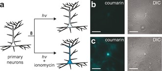
Figure 2.

Function of Ca2+‐sensitive photocaged coumarin in neurons. a) Representation showing the experimental setup. b, c) Fluorescence (left) and differential interference contrast (DIC) microscopy images (right) of live cultured hippocampal neurons incubated with AM ester 8 (10 μm) for 1 h and then illuminated with light of wavelength λ=365 nm for 20 s. Scale bars=100 μm. b) Untreated neurons. c) Neurons treated with ionomycin.
