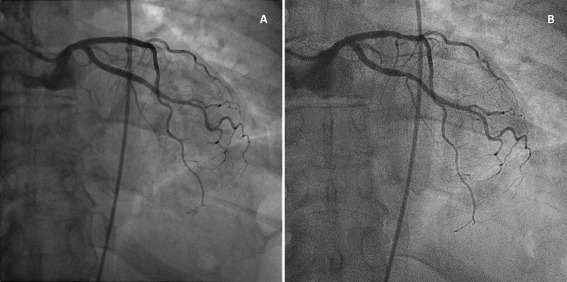
Figure 2.

A: Posterior‐anterior view of the LCA of a patient, acquired with the reference system (Room A). B: Posterior‐anterior view of the LCA of the same patient, acquired with the new imaging technology at 30% of the radiation dose (Room B). These images were acquired when the patient returned to the department for therapeutic intervention on a different day. Original moving images are available in online Supporting Information.
