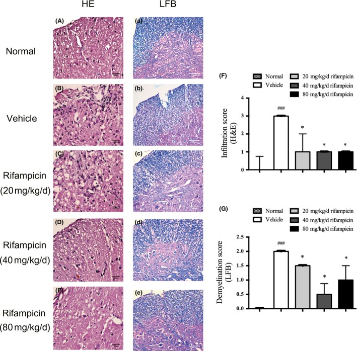
Figure 2.

Preventive rifampicin administration attenuates neuropathology of experimental autoimmune encephalomyelitis (EAE). Transverse sections of MOG 33‐35‐immunized mice treated with rifampicin or phosphate‐buffered saline (PBS) (n = 4 per group) starting from day 1 post‐immunization were stained to assess the level of neuropathology. Representative images are shown. The normal mice were used as a negative control (n = 4). (a–e) H&E staining. Inflammatory cell infiltration decreased remarkably in the rifampicin group compared with the vehicle group (bars = 500 μm) (a–e) luxol fast blue (LFB) staining. MOG induction caused severe demyelination that was significantly decreased by rifampicin treatment (bars = 80 μm). The cells in the infiltrates (f) and the demyelination region (g) were quantified with ImageJ software. The median infiltration and demyelination score of normal mice was 0, which bars are not shown in the histogram. Statistical analysis was performed using Mann–Whitney U test followed by Bonferroni post hoc test to compare replicate by time. Values are shown as median and upper limit (.PR75 quantile). Statistical significance: ###p < 0.001, compared with normal mice; *p < 0.05, compared with vehicle‐treated mice.
