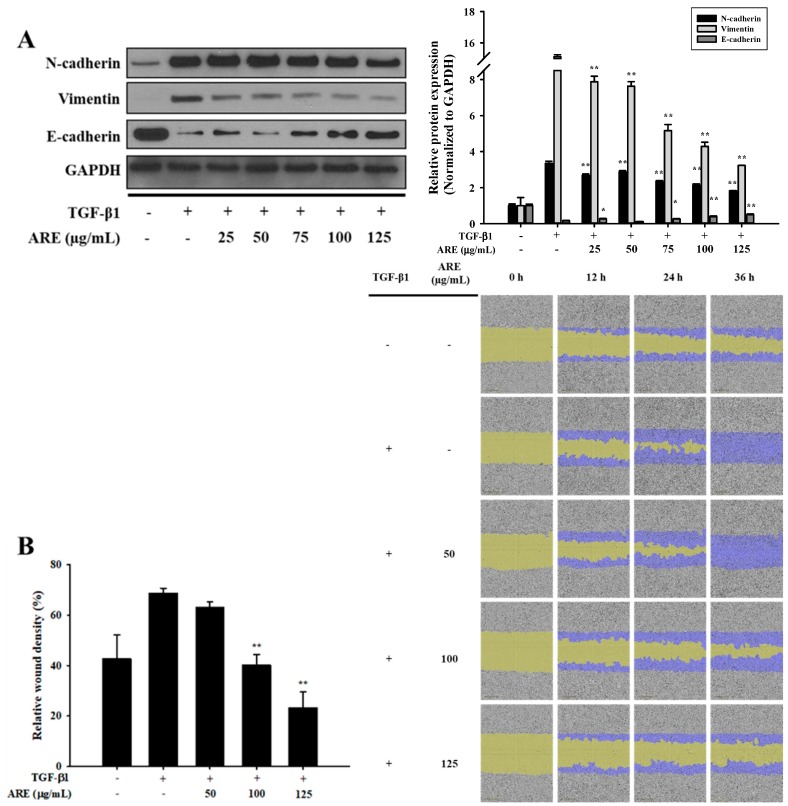
Figure 1.
Astilbe rubra extract inhibited TGF-β1-induced epithelial-to-mesenchymal (EMT) in A549 cells. A549 cells were co-treated with TGF-β1 and A. rubra extract (ARE) for 48 h. (A) The expression of N-cadherin, vimentin, and E-cadherin proteins were analyzed using Western blot analysis, with GAPDH expression used as a loading control. ImageJ was used for the quantification of the Western blots. (B) A549 cells were plated into 96-well plates and treated with TGF-β1 (5 ng/mL) or co-treated with ARE. The cell mobility was analyzed by IncuCyte ZOOM over 36 h at 10× magnification through a phase-contrast objective lens. The bar graph represents the wound density at 24 h exposure. All data are shown as mean ± S.D., n = 3. ** p < 0.01 vs. the TGF-β1-treated group.

