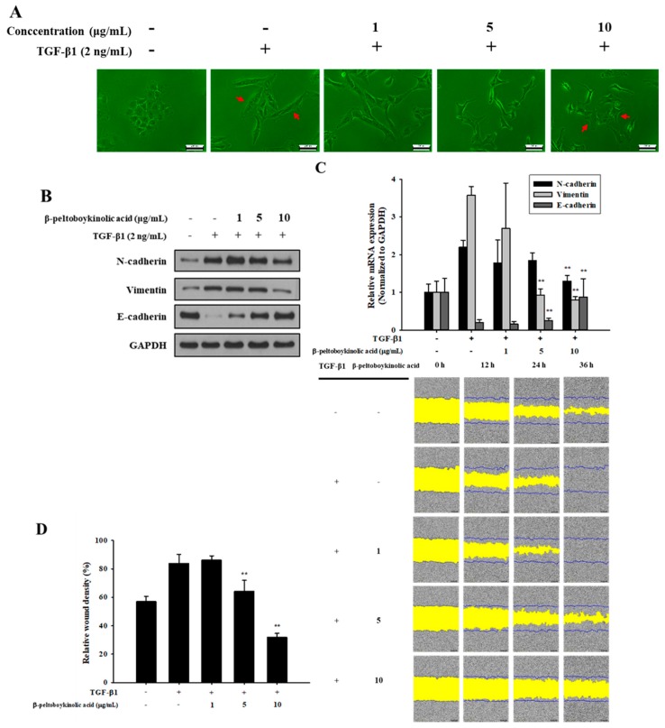
Figure 4.
Inhibitory effect of β-peltoboykinolic acid on TGF-β1-induced epithelial-to-mesenchymal (EMT) in A549 cells. A549 cells were treated with TGF-β1 or co-treated with β-peltoboykinolic acid for 48 h. (A) The morphology of A549 cells was observed using a phase-contrast microscope. Arrows indicate the difference between cell morphologies. Scale bar = 200 μm. (B) The expression of N-cadherin, vimentin, and E-cadherin proteins were analyzed using Western blot analysis, with GAPDH expression used as a loading control. (C) A549 cells were treated with TGF-β1 (2 ng/mL) or co-treated with β-peltoboykinolic acid (1, 5, and 10 μg/mL) for 24 h. The expression of N-cadherin, vimentin, and E-cadherin mRNA were analyzed using qRT-PCR. Fold change was calculated using 2−ΔΔCt relative quantitative analysis. (D) A549 cells were treated with TGF-β1 (5 ng/mL) or co-treated with β-peltoboykinolic acid (1, 5, and 10 μg/mL). The cell mobility was analyzed by IncuCyte ZOOM over 36 h at 10× magnification through a phase-contrast objective lens. The bar graph represents the wound density at 24 h exposure. All data are shown as mean ± S.D., n = 3. ** p < 0.01 vs. the TGF-β1-treated group.

