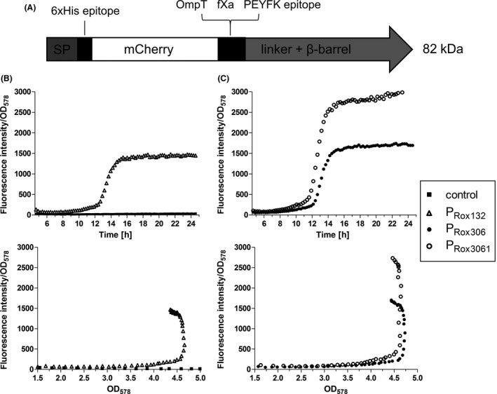
Figure 3.

A. Scheme of the unprocessed MATE‐mCherry fusion protein. After cleavage of the signal peptide (SP), the protein consists of a 6xHis epitope, the passenger mCherry, OmpT and fXa cleavage sites, a PEYFK epitope and the EhaA linker and β‐barrel (Sichwart et al., 2015).
B,C. Expression of MATE‐mCherry under control of PRox132, PRox306 and PRox3061. P. putida without plasmid and with plasmid pPRox132‐MATEmCherry (B), pPRox306‐MATE‐mCherry and pPRox3061‐MATE‐mCherry (C) were cultivated in LB medium in a 24‐well MTP at 30°C. OD 578 and fluorescence intensity (FI, excitation: 580 nm, emission: 620 nm) were monitored. These data are mean values of biological triplicates. Error bars are not visible due to small standard deviations that are covered by the icons.
