Figure 4.

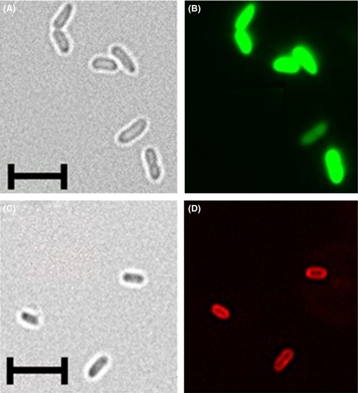
Analysis of P. putida pPRox3061‐sfGFP (A,B), and pPRox132‐MATE‐mCherry (C,D) via fluorescence microscopy. The strains were cultivated at 30°C, 200 rpm in LB medium for 8 h and 24 h respectively. 2.5 × 10⁶ cells were washed three times with PBS and fixed on a microscope slide with DABCO/Mowiol. The samples were analysed with the 100× oil immersion lens of a BZ‐9000 fluorescence microscope (Keyence, Neu‐Isenburg, Germany).
A,C. Brightfield pictures. (B) GFP‐filter, excitation 472/30 nm, emmission 593/40 nm.
D. TexasRed‐filter, excitation 560/40 nm, emmission 630/75 nm. The length of the scales corresponds to 5 μm.
