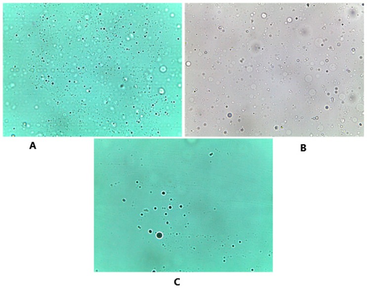
Figure 6.
Optical microscope images that show the spherical shape of the formed niosomes after hydration, and the distribution of these niosomes. The three different carriers were examined under the microscope accordingly. (A) Glucose niosomes (FN1) Magnificatin (Mag) = 20 X, (B) maltodextrin niosomes (FN2) Magnification (Mag) = 20 X, (C) mannitol niosomes (FN3) Magnification (Mag) = 20 X.

