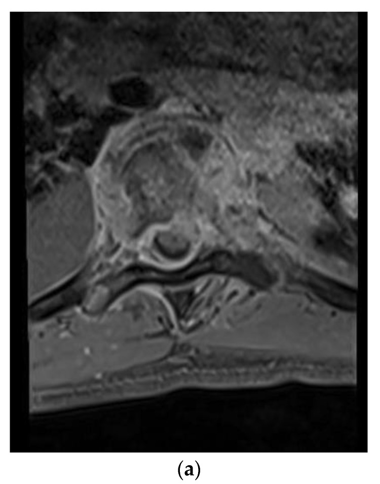
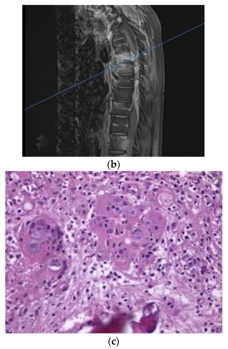
Figure 1.
(a) Axial T1-weighted magnetic resonance (MR) images with contrast showing extensive vertebral body and soft tissue enhancement with compression of the spinal canal at T6. (b) Sagittal T1-weighted MR Images with contrast showing extensive enhancement throughout the vertebral bodies and soft tissue but most significantly at T6–7. (c) Hematoxylin and eosin (H&E) staining of the thoracic bone specimen showing acute osteomyelitis with abundant coccidioides organisms. Multinucleated giant cells with engulfed coccidioides spherules is a characteristic finding. Abundant acute inflammatory changes was noted in the marrow cavity.


