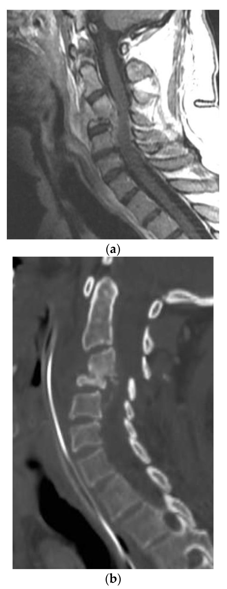
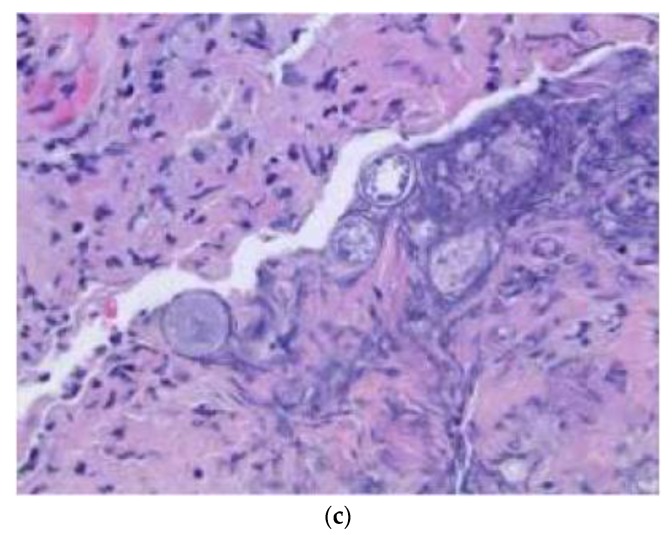
Figure 3.
(a) Sagittal T1-weighted MR images with contrast showing extensive disc space destruction at C3-4 with epidural enhancement. (b) Sagittal cervical spine CT showing postoperative erosion of the C4 vertebral body significant retrolisthesis of C3 onto C4. (c) H&E stain of the cervical spinal pathologic specimen demonstrating the Coccidioides spherules within soft tissue, which is morphologically compatible with coccidioidomycotic osteomyelitis.


