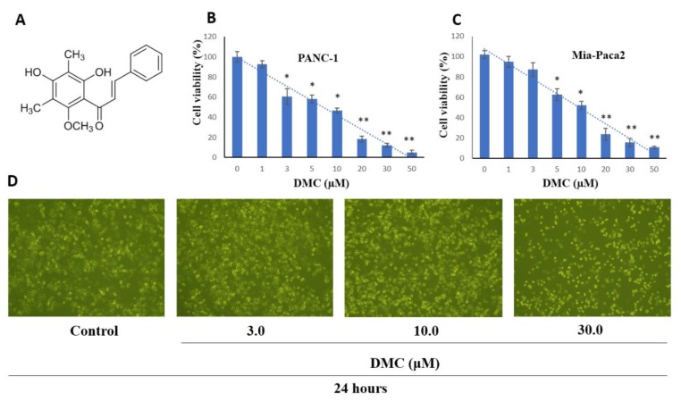
Figure 1.
(A) Chemical structure of DMC; Effect of DMC on PANC-1 (B), and MIA-PACA2 (C) cell viability; and (D) PANC-1 cell morphology visualized by light microscopy (scale bar 500 µm), cells were seeded into 6-well plates at 1 × 10⁵ cells/well and treated with the indicated concentration of DMC for 24 h. Data are presented as the mean ± standard deviation of three independent experiments performed in duplicate (*p < 0.01; **p < 0.05).

