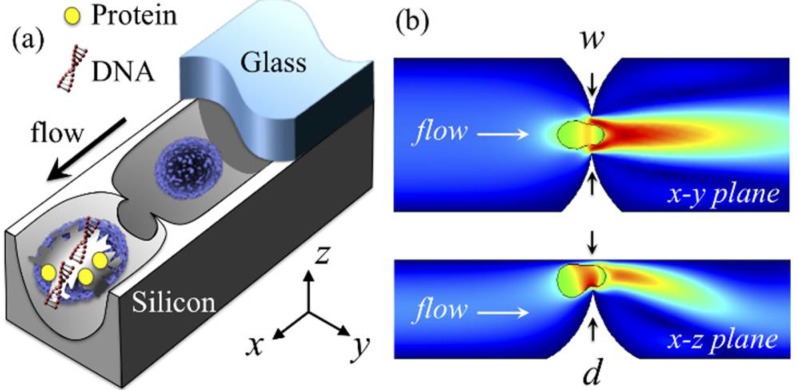
Figure 1.
Single-cell point constriction for reagent-free cell lysis. (a) 3D rendering: cell being ruptured by the ultra-sharp edge of a round constriction. (b) Cutaway views: cell undergoing excessive rapid deformation through a point constriction. Colour spectrum: flow velocity (increasing from blue to red). The glass cover is partially illustrated for clarity.

