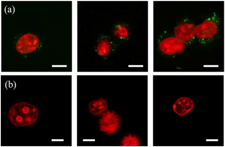
Figure 6.
Confocal fluorescence images of the nuclei (red; propidium iodide (PI)-stained) from mouse embryo fibroblast (NIH/3T3) cells isolated using (a) an eight-constriction treatment and (b) chemical lysis. The nucleus, and the nuclear doublet and triplet shown in (a) carry membrane fragments (green; DiOC18(3) (DiO)-stained). The constrictions are μm and μm. Scale bars are 10 μm.

