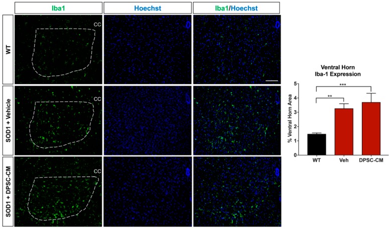
Figure 6.
DPSC-CM therapy in the late pre-symptomatic period did not significantly alter lumbar ventral horn microglial reactivity. Microglial reactivity as measured by percent area of the ventral horn (dashed white line) occupied by Iba1+ labeling (green) was not significantly different between DPSC-CM and vehicle treated mSOD1G93A mice at PD91. As with reactive astrogliosis, both treatment groups exhibited a similar significant elevation of microglial reactivity compared to WT mice. Data represent mean +/− SEM. Data analyzed via one-way ANOVA. (WT, n = 3; mSOD1G93A, n = 5–6). **, p < 0.01; ***, p < 0.001. CC = central canal. Scale bar = 100 μm.

