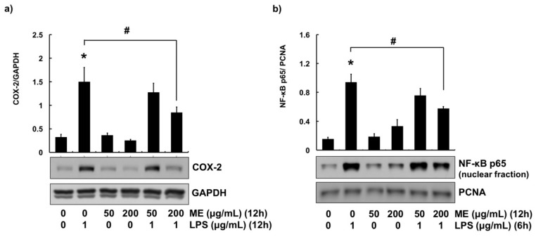
Figure 3.
COX-2 expression and NF-κB activation in cells treated with moringa extract (ME). (a) Cells were pre-treated with ME (50 and 200 µg/mL) for 12 h followed by LPS treatment (1 µg/mL) for 12 h. Western blot analysis was used to measure COX-2 expression levels in whole cell lysates. GAPDH was used as a loading control. (b) Cells were pre-treated with ME (50 and 200 µg/mL) for 12 h followed by LPS treatment (1 µg/mL) for 6 h. Nuclear translocation of NF-κB p65 was determined using nuclear fraction and Western blot analysis. PCNA was used as a loading control. Western blots shown are representative images of three independent experiments. Values represent means ± SEM (n = 3). *, significant difference vs. control (p < 0.05). #, significant difference vs. LPS only (p < 0.05).

