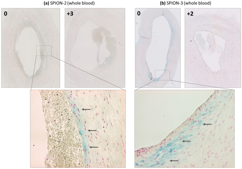
Figure 3.
Detection of iron presence in arterial wall by histology. Magnetically targeted (segment 0) and representative control segments away from the tip of the magnet are shown for (a) SPION-2 and (b) SPION-3, suspended in the whole blood. Arrows indicate blue staining due to the particle accumulation.

