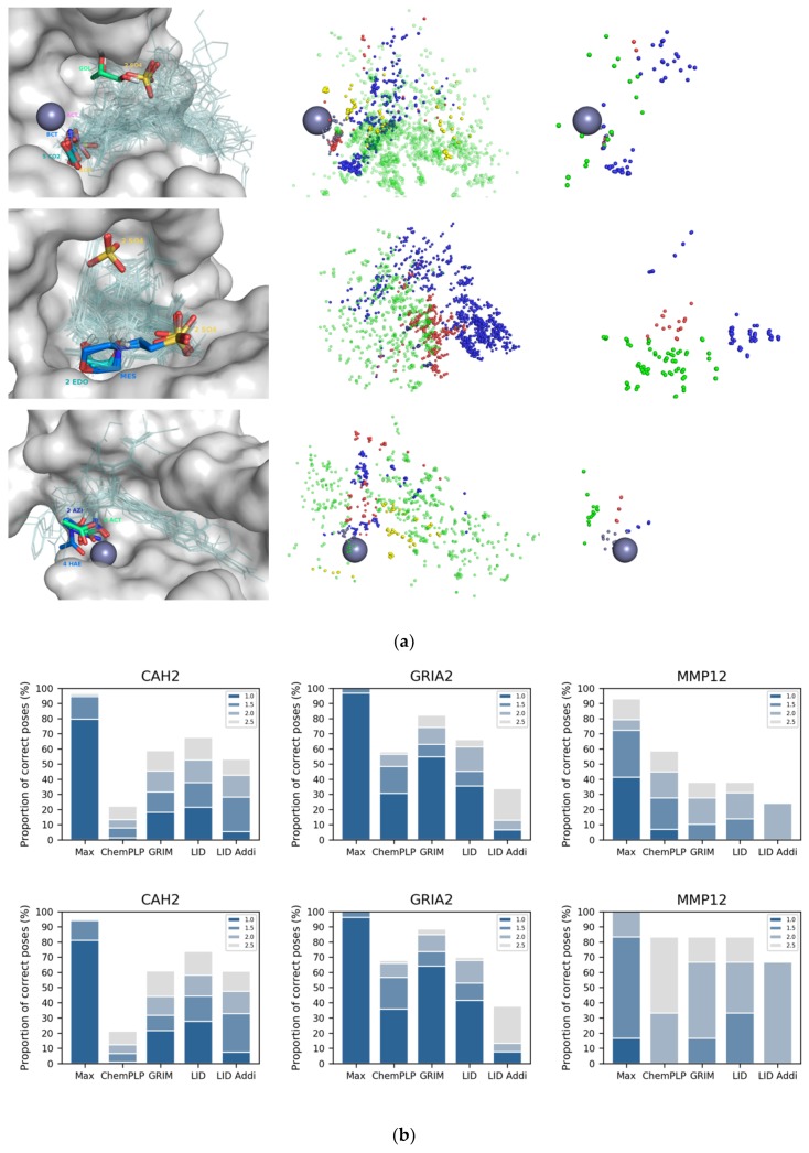
Figure 4.
Use of crystallization additives binding modes in LID. (a) additives crystallized in CAH2 (top), GRIA2 (middle) and MMP12 (bottom). Metal cation is represented with a grey sphere. On the left triad are shown the additives (thick sticks colored by HET code) and drug-like ligands (transparent lines) in the protein site (grey surface); on the middle triad are shown the drug-like ligands interaction pseudo-atoms, colored according to the corresponding bond; on the right triad are shown the additives interaction pseudo-atoms, colored according to the corresponding bond type (hydrogen bond in red and blue, π-stacking in yellow, hydrophobic contact in green and metal chelation in grey); (b) LID’s performance in pose prediction of drug-like ligand (top) and fragment only (bottom). LID and LID Addi refer to the use of drug-like ligands and additives as reference, respectively.

