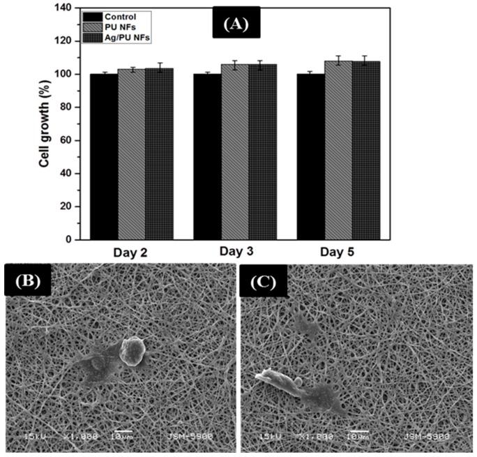
Figure 8.
NIH-3T3 cell proliferation on various nanofiber membranes after culture for one, three, and five days (A). The viability of the control cells was set to 100% and the viability relative to the control was expressed. (B,C) represent the SEM images of the cell fixation test on the PU NFs and Ag/PU NFs after five days of incubation, respectively.

