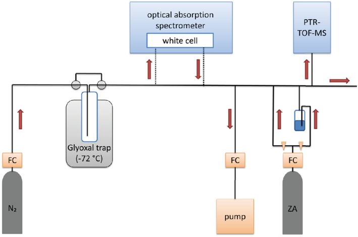
Figure 1.

Experimental setup for the calibration of glyoxal. In the upper part of the figure are the UV absorption spectrometer and the Proton Transfer Reaction Time of Flight Mass spectrometer (PTR‐TOF‐MS). The lower part shows the glyoxal source in a dry ice‐ethanol bath at −72 °C, the pump with a flow controller (FC) to reduce the sample flow and the dilution setup with a zero air gas bottle (ZA) and two tubes in order to adjust the humidity with a bubbler. The red arrows represent the flow direction.
