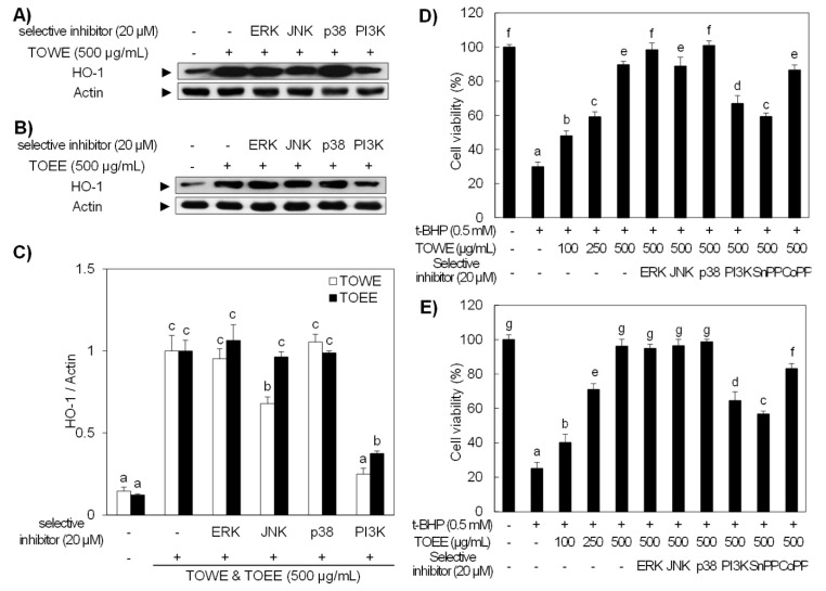
Figure 4.
TOWE and TOEE induced HO-1 expression was abolished by the treatment of selective inhibitors, which was confirmed by t-BHP-induced oxidative damage in RAW 264.7 cells. (A,B) RAW 264.7 cells were treated with 20 μM of selective inhibitors for PI3K/Akt and MAPK signaling molecules in the presence of 500 μg/mL of TOWE and TOEE. The selective inhibitors for JNK and PI3K attenuated TOWE-induced HO-1 expression, while TOEE-induced HO-1 expression was only abolished by a PI3K selective inhibitor. The HO-1 protein expression was analyzed by Western blot analysis. (C) The relative induction of HO-1 was quantified by densitometry, and actin was used as an internal control. (D,E) The antioxidative potential of TOWE and TOEE scavenged the t-BHP-induced oxidative damage in RAW 264.7 cells. RAW 264.7 cells were treated with various concentrations of TOWE or TOEE for 12 h in the presence or absence of each selective inhibitor or inducer. The cells except untreated were exposed to 0.5 mM t-BHP for 3 h. The data represent the mean ± standard deviation of triplicate experiments. The values sharing the same superscript are not significantly different at p < 0.05 by Duncan’s multiple range test. t-BHP, tert-butyl hydroperoxide; SnPP, tin protoporphyrin; CoPP, cobalt protoporphyrin; PI3K, phosphoinositide 3-kinase.

