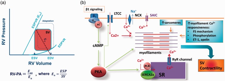
Fig. 3.
(a) Schematic illustration of pressure-volume relationship of the right ventricle (RV) and RV–pulmonary artery (PA) coupling. An adapted RV pressure–volume relationship is also shown (defined by dotted square). Ea, arterial elastance; EDPVR, end-diastolic pressure-volume relationship; EDV, end-diastolic volume; ESD, end-systolic volume; ESPVR, end-systolic pressure-volume relationship, defined as contractility and known as Ees; SV, stroke volume. (b) Mechanism by which contractility is maintained in adapted or compensated RV during PH. An increase in afterload activates three fundamental mechanisms that increase contractility within the RV. These mechanisms remain active throughout the adaptive/compensated phase. See text for details. AC, adenylate cyclase; FS, Frank-Starling; ET-1, endothelin 1; LTCC, L-type calcium channel; PKA, protein kinase A; RyR, ryanodine receptor; SAIC, stretch-activated ion channels; SR, sarcoplasmic reticulum.

