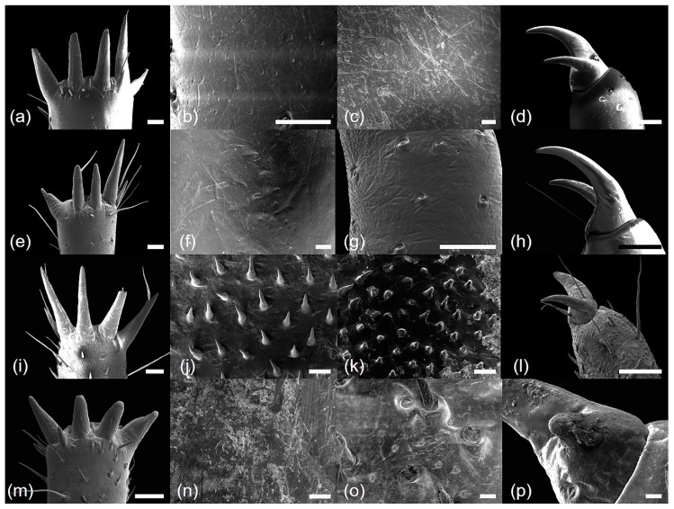
Figure 5.
Scanning electron microscope micrographs of four hind leg parts: (a–d) Cryptotympana atrata; (e–h) Hyalessa maculaticollis; (i–l) Meimuna opalifera; (m–p) Platypleura kaempferi; (a,e,i,m) spine of metatibia; (b,f,j,n) surface of metatibia; (c,g,k,o) surface of metatarsus; (d,h,l,p) apex of metatarsus. All scale bars = 200 μm (except b, c, f, j, k, n, o, p, scale bars = 20 μm).

