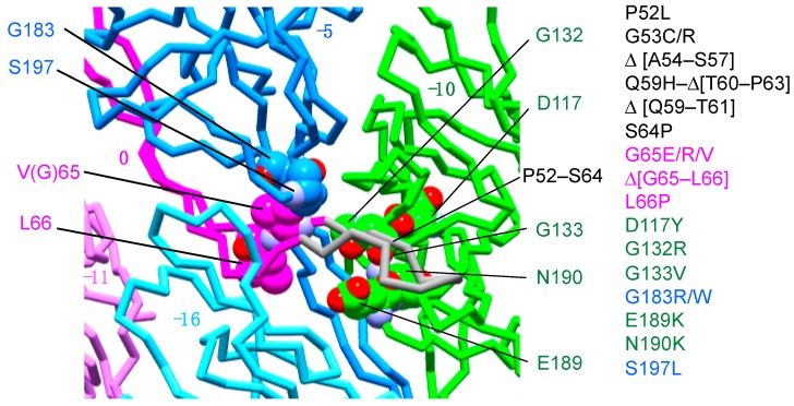
Figure 4.
Polyrod mutation sites are localized around the tip of the l-stretch. The known polyrod mutation sites are mapped on the rod structure. Subunit at 0, −5, −10 positions are colored in magenta, blue, and green, respectively. The putative structure of the l-stretch tip of subunit 0 is shown in gray. The polyrod mutation sites except for those in the l-stretch tip are indicated by ball models. Oxygen and nitrogen atoms are colored in red and purple, respectively. The carbon atoms are painted by the same color as used for the subunits. The polyrod mutations [32] are listed to the right of the figure. Molecular figures were drawn using MolFeat (Ver 3.6, FiatLux Corporation).

