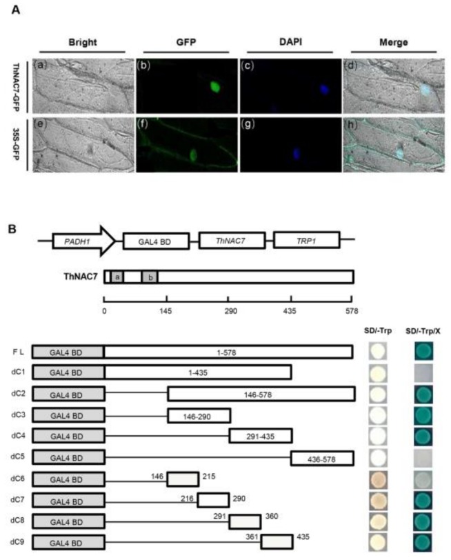
Figure 2.
Subcellular localization and transcriptional activation of ThNAC7. (A): Subcellular localization analysis of ThNAC7. The onion nuclei were visualised through DAPI (4′,6-diamidino-2-phenylindole) staining. Bars, 50 μm. (B): Transactivation assay of ThNAC7 (a, b: NAC domain). A diagram of the pGBKT7 constructs for expressing different truncated ThNAC7 proteins in yeast cells. Transactivation assay of the intact or truncated ThNAC7 proteins. Full length or truncated CDSs of ThNAC7 were constructed into pGBKT7 vector and transformed into Y2HGold cells, and grown on SD/-Trp or SD/-Trp/-His/X-α-gal mediums to assess their transcriptional activation.

