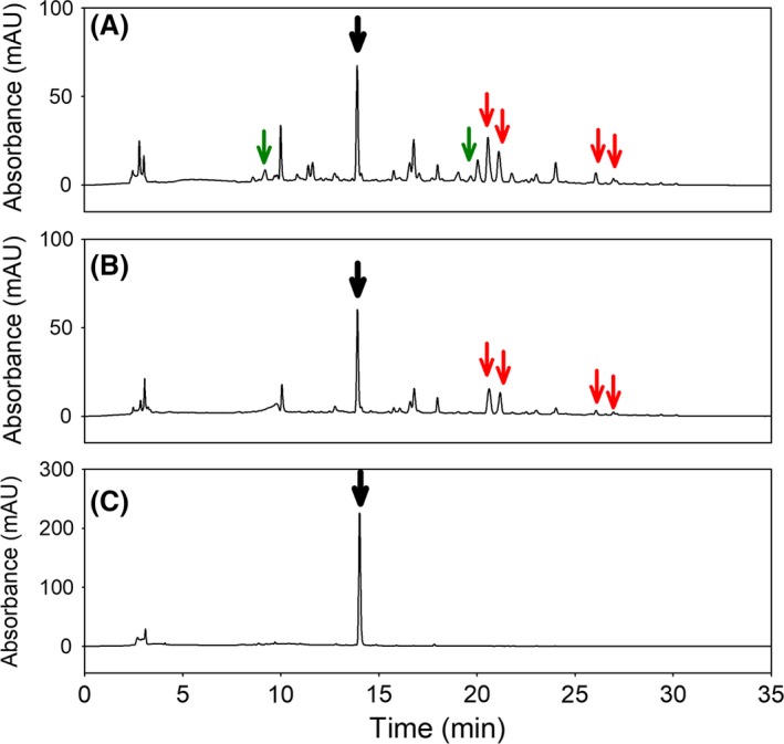
Figure 3.

Production of dopaxanthin in microbial cultures. Chromatograms at λ = 480 nm for the detection of betaxanthins in the media LB (A), NZCYM (B) and water (C). Dopaxanthin was detected at 13.9 min with a signal intensity higher in water than in LB and NZCYM (black arrows). Betaxanthins present exclusively in LB medium are indicated with green arrows. Red arrows indicate betaxanthins present in both LB and NZCYM media.
