Figure 6.
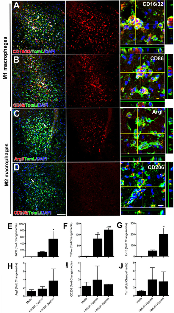
Intraspinal injection of recombinant HMGB1 (500ng in 1μl) elicits neuroinflammation dominated by neurotoxic inflammatory M1 microglia/macrophages. Three days after injection, a robust CNS macrophage response is evident in the ventral horn (A-H). Immunohistochemical staining for classical M1 macrophage activation markers (CD16/32 & CD86) reveals that most macrophages exhibit an M1 phenotype (A-B). A few cells express M2 macrophage markers (C-D). In vitro, BMDMs treated with recombinant HMGB1 increase expression of M1 markers (E-G), but not M2 markers (H-J). (ANOVA; *p<0.05, **p<0.01, ***p<0.001; Scale = 100μm (10μm for orthogonal view).

