Figure 2.
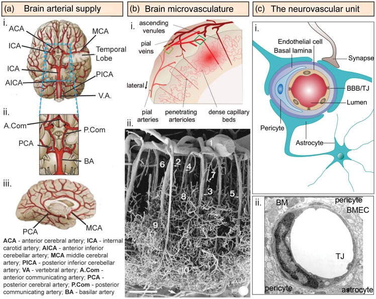
Cerebrovascular architecture. (a) Arterial architecture: (i) inferior view of the base of the brain with cerebral arterial Circle of Willis; (ii) magnified view of the Circle of Willis; and (iii) right lateral view of the right hemisphere. (b) Microvasculature ultrastructure in the cerebral cortex: (i) schematic illustration of the vasculature in the cerebral cortex showing both arterial and venous systems. Pial arteries located on the surface penetrate deep into the cerebral cortex as penetrating arterioles, which branch into capillary beds that then reemerge from the cortex as ascending venules.14 (ii) Scanning electron micrograph of a corrosion cast showing the vasculature of the temporal lobe of the human cerebral cortex.16 (Scale bar = 375 µm): (1) pial artery, (2) long cortical artery, (3) middle cortical artery, (4) short cortical artery, (5) cortical vein, (6) subpial zone, (7) precapillary vessel with blind ending, (8) superficial capillary zone, (9) middle capillary zone, and (10) deep capillary zone. (c) Neurovascular unit. (i) Schematic illustration of the neurovascular unit comprised of brain microvascular endothelial cells surrounded by pericytes and astrocytes.85 (ii) Electron microscope cross section of a capillary from the rat frontoparietal cortex.163
BM: basement membrane; BMECs: brain microvascular endothelial cells; TJ: tight junction.
Source: Figure 2(b)(i) is reproduced with permission from Chen et al.;14 Figure 2(b)(ii) is reproduced with permission from Reina-De La Torre et al.;16 Figure 2(c)(i) is reproduced with permission from Walchli et al.;85 and Figure 2(c)(ii) is reproduced with permission from Farkas & Luiten.163

