Figure 3.
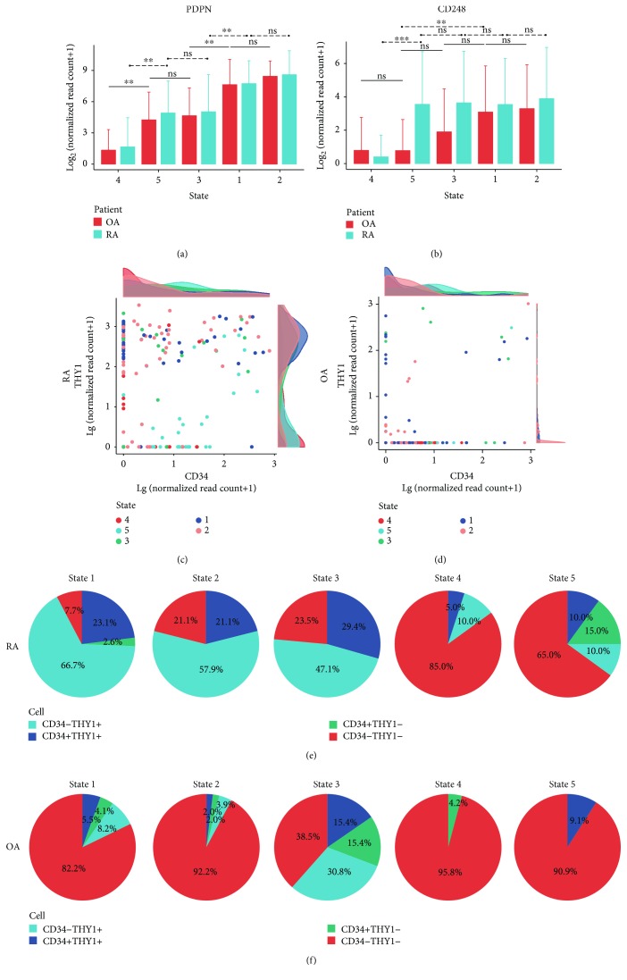
Prediction of transition orientation and localization of SFs. Average expression level (log2 transformed) of PDPN (a) and CD248 (b) in SFs of states 1 to 5. OA SFs: red; RA SFs: blue. Expression level of CD34 and THY1 in RA (c) and OA (d) SFs. Plots represent SFs. Density plots show the distribution of expression level (log10 transformed) of CD34 (upper) and THY1 (right) in the corresponding plot. Different colors represent different states. Composition of cell subsets divided by CD34 and THY1 across states 1 to 5 in both RA (e) and OA (f) SFs. Blue: CD34-THY1+; dark blue: CD34+THY1+; green: CD34+THY1-; red: CD34-THY1-. CD34+/THY1+: lg(normalized read counts of CD34/THY1 + 1) ≥ 1.5. Bars show means, and error bars show standard deviations. ∗ p < 0.01, ∗∗ p < 0.001, and ∗∗∗ p < 0.0001; ns = not significant.

