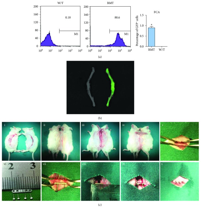
Figure 1.
Bone marrow transplantation (BMT) and the surgical procedures of complex animal models. (a) At 6 weeks, GFP+ cells in bone marrow of BMT and wild-type mice were detected by flow cytometry. The numerical value represents the mean percentage of GFP+ cells in each group (n = 3). (b) Bioluminescence image of femur and tibia. Green fluorescence intensity was observed between wild-type (left) and BMT (right). (c) The parabiotic mouse model was fabricated (i-iv) and two weeks later, a critical-sized bone defect was created (v-x).

