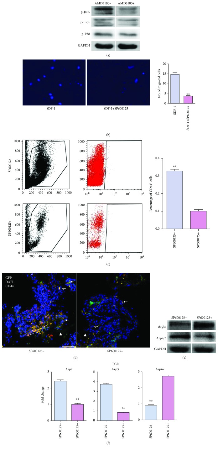
Figure 5.
JNK served as an effector downstream of SDF-1/CXCR4 in the migration of bone marrow (BM) CD44+ cells towards tissue-engineered constructs (TECs). (a) Comparison of phosphorylated JNK (p-JNK), ERK (p-ERK), and P38 (p-P38) expression in BM cells after CXCR4 blockade. After migration stopped, BM cells were collected and analyzed by western blot. (b) Representative images of cell migration towards SDF-1. The quantification of migrated BM cells is shown as a bar graph (n = 5). ∗∗P < 0.01. (c) FACS analysis of the proportions of CD44+ cells in peripheral blood (PB). The quantification comparison is shown as a bar graph (n = 3). ∗∗P < 0.01. (d) Representative images of in vivo migration of GFP+/CD44+ cells towards TECs. The introduction of SP600125 significantly reduced the recruitment of GFP+/CD44+ cells. White triangle, implant area; white arrows, CD44+ cells; scale bar, 50 mm. (e) Comparison of Arpin and Arp2/3 expressions in BM cells after JNK blockade in vitro. (f) Analysis of the Arpin, Arp2, and Arp3 mRNA expression in sorted CD44+ cells. On day 3 postoperatively, cells in PB were sorted by FACS on CD44. RT-PCR was performed to evaluate Arpin, Arp2, and Arp3 mRNA expression. The quantification data are shown as a bar graph (n = 3). ∗∗P < 0.01.

