Figure 8.
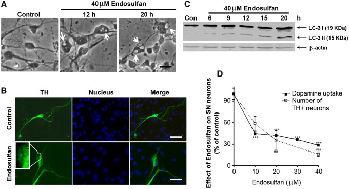
Endosulfan-induced autophagy and dopaminergic neuronal cell loss in primary mesencephalic cultures. (A) Primary cultures of striatal (STR) neurons were treated with 40 μM endosulfan for 12 or 20 h. Scale bar, 10 μm. Autophagic vacuoles were examined by phase-contrast microscopy. (B) Mesencephalic primary cultures were treated with 40 μM endosulfan for 20 h and morphological changes were examined, including autophagic vacuole formation. Scale bar, 10 μm. TH staining was used to define the TH+ dopaminergic neurons. (C) Immunoblot analyses of accumulating LC3-II protein in STR neurons treated with 40 µM endosulfan. β-actin expression was used as an equal loading control. (D) Assessment of viability of dopaminergic neurons using 3H-DA uptake assay and the number of TH+ neurons. Asterisks (##, p < .01, *** and ###, p < .001) denote significant treatment effects relative to the vehicle-only dose (0 µM endosulfan).

