Fig. 1.
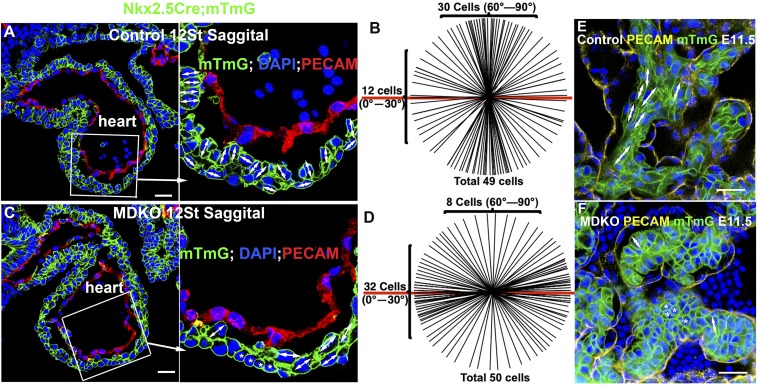
NFPs are required for cellular orientation and organization during cardiac morphogenesis. Most of cardiomyocytes in the inner layer of the compact zone from a control heart orient perpendicularly to the heart wall (A and B), while most of the cells in the MDKO display no or parallel orientation to the heart wall (C and D). The double-headed arrows indicate the orientation of a cell and the asterisk indicates no orientation of a cell in A and C. B shows the cellular orientation of 49 cells of control hearts and D shows 50 cells in MDKO hearts. Each line represents an orientation of a cell in the inner layer in A or C and the red line represents the heart surface reference line in B and D. (E and F) The cells in control trabeculae display a long spindle shape and align parallel to trabecula (E), while cells in MDKO trabeculae do not show orientation and are round, indicated by an asterisk (F). (Scale bars in A, C, E, and F: 20 μm.)

