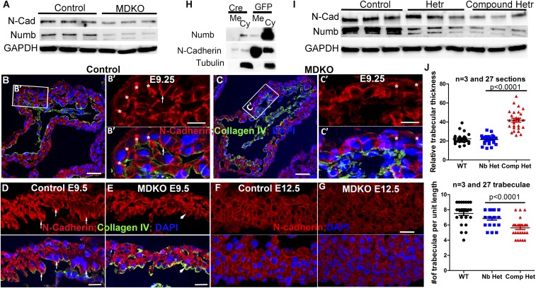
Fig. 4.
NFPs regulate trabecular morphogenesis through N-cadherin. (A) MDKO hearts display lower levels of Numb and N-cadherin than control hearts at E13.5. (B) N-cadherin localizes to the lateral domain of the cardiomyocytes in the myocardium at E9.25, indicted by the asterisk, and to the lateral and apical domains of some cardiomyocytes that face toward the heart lumen, indicated by the arrow. (C) N-cadherin localization to the lateral domain is weaker in the MDKO, as indicated by the asterisk. (D and E) N-cadherin localizes to the apical domain strongly in the control heart at E9.5 but weakly in the MDKO heart. The weaker membrane localization in the MDKO is further demonstrated in E12.5 hearts. (F and G) The membrane fractionation shows that the membrane N-cadherin in cultured NFP null cardiomyocytes is significantly reduced compared with the controls (H). (I) Levels of Numb and N-cadherin proteins are reduced in the compound heterozygotes. (J) Compound heterozygous hearts (Nkx2.5Cre/+;Nbfl/fl; Nlfl/+;Cdh2fl/+ or Nkx2.5Cre/+; Nbfl/+; Nlfl/fl; Cdh2fl/+) display thicker and less-dense trabecula compared with the single heterozygotes, which are not significantly different from control. (Scale bars in B, C, F, and G: 20 μm and in D and E: 10 μm.)

