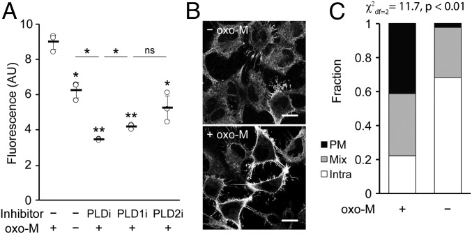
Fig. 7.
Muscarinic M1 receptor (M1R) activation leads to PLD activity that is predominantly at the plasma membrane. HeLa cells stably expressing M1R were labeled for RT-IMPACT by treatment with the indicated PLD inhibitor or DMSO for 30 min (A only) and then simultaneous treatment of (S)-oxoTCO–C1 in the presence or absence of oxo-M (5 min), rinsing (1 min), and IEDDA reaction with Tz–BODIPY (0.33 µM) for 1 min followed by flow cytometry analysis (A), with mean fluorescence intensity in arbitrary units (AU) indicated, or (B and C) in real time with time-lapse confocal images taken (B) and quantified (C) 9 s after the addition of Tz–BODIPY, using the PM/Mix/Intra rubric described in Fig. 5. In B, the brightness of the –oxo-M image was increased to facilitate comparison of the localization of IMPACT-derived fluorescence in the 2 images. (Scale bars: 20 µm.) For A, statistical significance was assessed by using 1-way ANOVA followed by Games–Howell post hoc analysis. Asterisks directly above data points (*P < 0.05 and **P < 0.01; ns, not significant) denote statistical significance compared with the first sample (−inhibitor, +oxo-M), and error bars represent SD. For C, statistical significance was assessed by using a χ2 test for independence, with the χ2 value (df, degrees of freedom) and associated P value indicated. Each bar contains data of 3 (−oxo-M) or 8 (+oxo-M) biological replicates, with n = 41 (−oxo-M) or 112 (+oxo-M) total cells.

