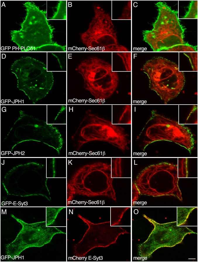
Fig. 1.
Assembly of ER-PM contact sites in HeLa cells expressing recombinant GFP-JPHs or GFP-E-Syt3. Live imaging of HeLa cells transfected with plasmids encoding the GFP-tagged PH domain of the PLC (GFP-PH-PLCδ1) (A), Junctophilin-1 (GFP-JPH1) (D and M), Junctophilin-2 (GFP-JPH2) (G), Extended Synaptotagmin-3 (GFP-E-Syt3) (J), or mCherry-tagged E-Syt3 (mCherry E-Syt3) (N), and Sec61β (mCherry-Sec61β) (B, E, H, and K). C, F, I, L, and O are merged images. Insets show higher magnification of selected regions of the PM of transfected cells. Scale bar, 5 μm.

