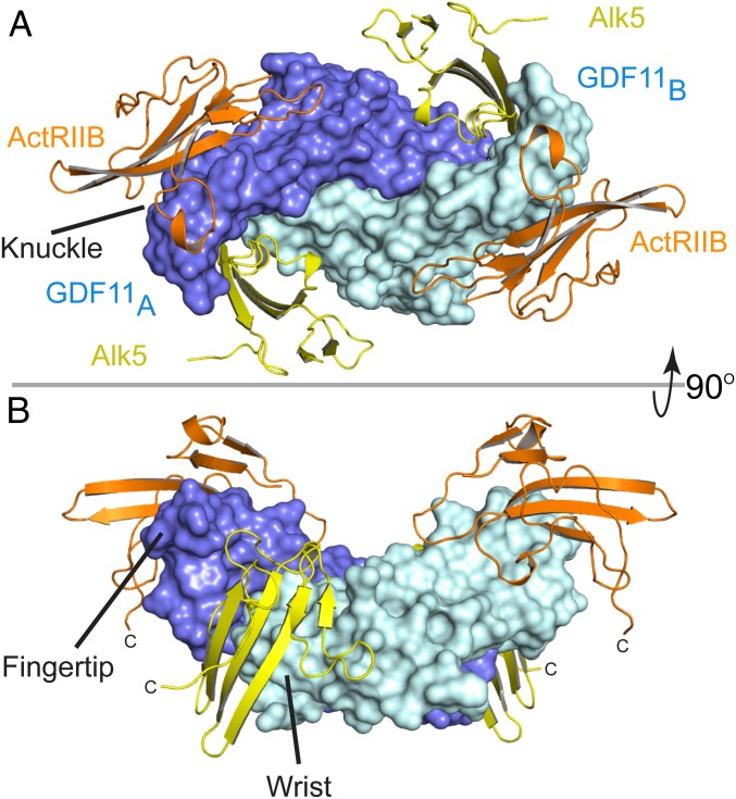
Fig. 1.
Structure of GDF11/ActRIIB/Alk5 ternary complex. (A) GDF11/ActRIIB/Alk5 as viewed on the membrane surface. GDF11 has 2 monomers represented in slate (monomer A) and cyan (monomer B). ActRIIB-ECD is represented in orange, with Alk5-ECD in yellow. (B) Ninety-degree upward view of the ternary complex. ActRIIB binds at the convex knuckle region of GDF11 whereas Alk5 binds at the concave interface of GDF11 formed between the fingertip of monomer A and the wrist helix of monomer B GDF11.

