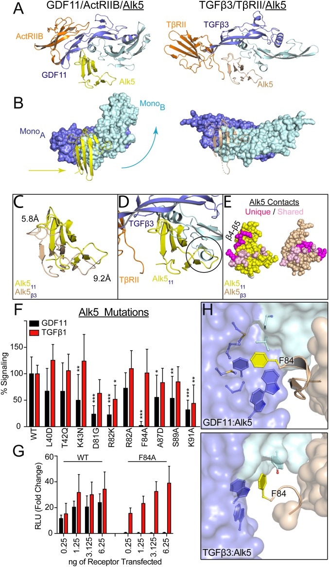
Fig. 3.
Differential binding of shared type I receptor Alk5 to GDF11 and TGFβ. (A) Ribbon showing 1 type I and 1 type II for GDF11/ActRIIB/Alk5 (PDB ID code 6MAC) and TGFβ3/TβRII/Alk5 (PDB ID code 3KFD), with orientation consistent with alignment of monomer A of the ligand (9). (B) Surface representation of GDF11 (Left) and TGFβ3 (Right) to highlight the relative positional differences of monomer B and Alk5. Arrows indicate the directional shift of GDF11 and Alk5 relative to TGFβ3. (C) Overlay of Alk5 bound to GDF11 (yellow) and TGFβ3 (sand), respectively. Alignment was performed using monomer A. Only the receptor is shown to highlight the shifts in both the β4–β5 loop and the N-terminal region. (D) Overlay of GDF11-bound Alk5 onto TGFβ3 complex using monomer A for superposition. Circle indicates a steric clash with the N terminus of the ligand that would occur if Alk5 were to bind TGFβ3 in a similar position as GDF11. (E) Surface interactions (within 5 Å) of Alk5 with both GDF11 and TFGβ3, with shared residues in pink and unique ligand-interacting residues in magenta. (F and G) Luciferase reporter assay in R1B L17 cells of GDF11 and TGFβ1 signaling following transient transfection of 1.25 ng of Alk5 variants and 2.5 ng (CAGA)12 promoter (F) or titration of Phe84-Ala (F84A) Alk5 transfection (G) (*P ≤ 0.05, **P ≤ 0.01, and ***P ≤ 0.001, 1-way ANOVA). (H) Differences in how F84 of Alk5 binds to GDF11 (Top) and TGFβ3 (Bottom).

