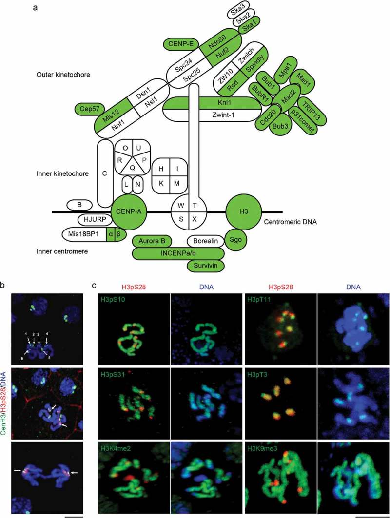
Figure 1.

O. dioica kinetochore and inner centromere complements and epigenetic marks. (a) Orthologs that were detected (green) in the O. dioica genome are projected onto the human complement (modified from [1]). Centromere proteins (CENPs) of the CCAN are represented by single letters. (b) Identification of O. dioica centromeres using CenH3. Top left: Six monocentric chromosomes in mitotic phase were indicated by numbers. Middle: CenH3-GFP (arrow) flanked H3pS28 signal on the centromeres. Bottom: CenH3-GFP localized at the centrosome proximal side of separating sister chromatids at anaphase. (c) The distribution of major histone H3 phosphorylation and methylation modifications on O. dioica chromosomes. H3pT3 and H3pS28 localized on the inner centromeres, and H3pT11 localized on the outer centromeres. In striking contrast, H3pS10, H3pS31, H3K4me2 and H3K9me3 localized on the chromosome arms. Bars, 3 µm.
