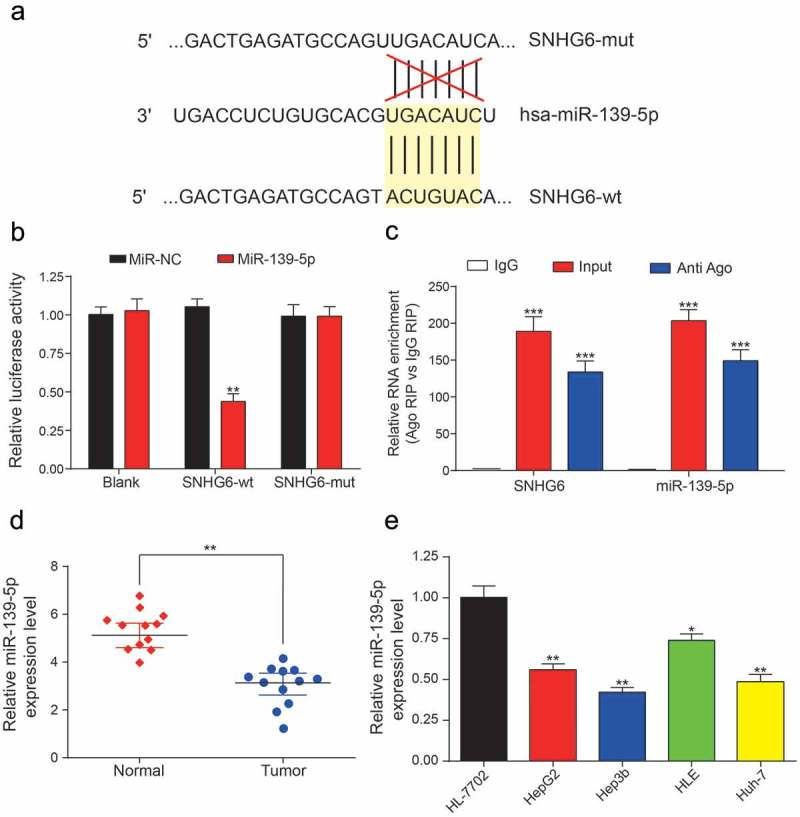
Figure 7.

Direct targeted relationship between SNHG6 and miR-139-5p. (a) The binding sites between wild type SNHG6/mutant SNHG6 and miR-139-5p. (b) Dual luciferase reporter gene assay verified that miR-139-5p could be targeted by SNHG6-wt rather than SNHG6-mut (*P< 0.05, compared with the miR-NC group) (c) MiR-139-5p and SNHG6 abundance in Ago-RIP fractions as measured by qRT-PCR. (d-e) MiR-139-5p expression was significantly lower in HCC tissues and cell lines compared with adjacent normal tissues and normal liver cell line, respectively. Expression levels were calculated from the gene/β-actin expression ratio (*P< 0.05, **P< 0.01, compared with the normal group or HL-7702 group). Every experiment was performed for 3 times at least.
