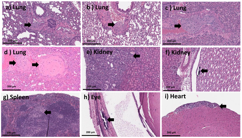
Figure 5.
20X H and E microscopic images from the organs of the mice which recovered from MSNPs500 (at dose range of ~200-389 mg.kg−1) (a-f, h-i) and Stöber SNPs50(at dose range of ~241-153 mg.kg−1) (g) 10 days post injection. This data shows lung tissue with organizing thrombosis involving larger pulmonary vessels (a-d), wedge shaped injury of the renal parenchyma, in keeping with infarction (e), focal calcifications inside the renal pelvis (f), spleen with aggregates of foamy histocytes (g), focal retinal calcifications (h), and focal calcified myocytes (i). Histologic abnormalities are marked with the black arrow.

