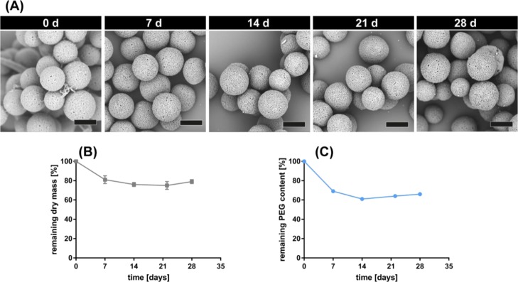
Figure 3.
In vitro degradation of placebo microspheres prepared with polymer A: polymer B blend of 50:50 after incubation at 37 °C in PBS pH 7.4, supplemented with 0.025% Tween 20, and 0.02% NaN3. (A) SEM images before and after incubation. Scale bars represents 30 μm. The time of incubation (d: days) is stated above each image, whereby “0 d” shows freshly prepared microspheres before incubation. (B) Remaining microsphere dry mass after 28 days and (C) remaining PEG content in the microsphere samples, as determined by 1H NMR.

