Figure 8.
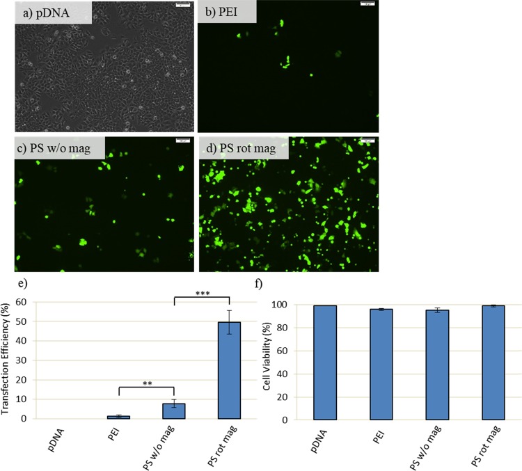
Transfection efficiency and viability assay at 3.5 cm distance and 1 h: MCF7 cells are transfected with 60 μg of PEI or PS together with 10 μg of GFP-DNA. Following transfection, cells are exposed to the magnetic field for 1 h and then washed with PBS right after magnetofection to remove uninternalized nanoparticles. Cells are incubated at 37 °C in fresh DMEM medium until incubation time is achieved to 48 h. (a–d) GFP expression is observed by inverted fluorescence microscopy at 48 h post-transfection. Scale bar: 100 μm. (e) Quantification of transfection efficiency analyzed by counting at least 900 cells for each condition. (f) Cell viability obtained by MTT assay. Cells are treated for 4 h with 0.5 mg/mL MTT in complete medium. Then, the absorbance of formazan solution is measured by using enzyme-linked immunosorbent assay. pDNA only, PEI, and PS w/o mag (PEI-SPION without magnetic field exposure) were used as control and treated in the same way as their counterparts. PS rot mag indicates PEI-SPION exposed to rotary magnetic fields. Data were shown as mean ± SD of at least 3 independent experiments; **p < 0.01, ***p < 0.001.

