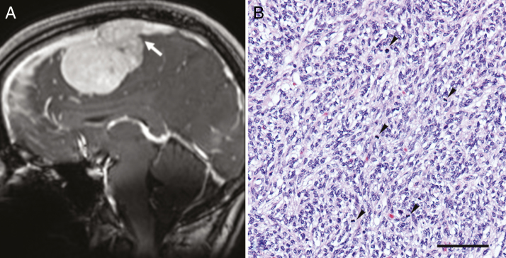
Fig. 1.
(A) Sagittal (coronal) post-contrast T1-weighted image shows avid enhancement in the lesion with invasion of the superior sagittal sinus (arrow). (B) The tumor consists of spindle cells with a vague fascicular-storiform pattern. The nuclei are ovoid and monomorphic, with frequent mitotic figures (arrowheads); hematoxylin and eosin; bar = 100 microns.

