Image 1.

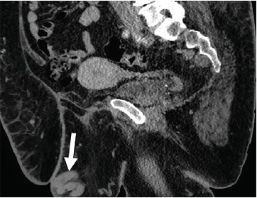
Sagittal cross-section of computed tomography of abdomen and pelvis with intravenous contrast demonstrating dilated pelvic veins (arrow) near the mons pubis.

Sagittal cross-section of computed tomography of abdomen and pelvis with intravenous contrast demonstrating dilated pelvic veins (arrow) near the mons pubis.