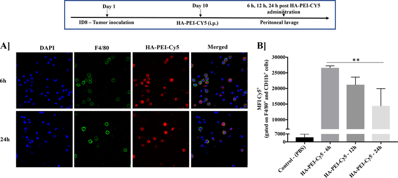
Figure 2. Uptake of HA-PEI-Cy5 nanoparticles by macrophages in ID8 tumor-bearing mice.
Female C57BL/6 mice were injected with ID8 cells on day 1. On day 10, the mice were injected intraperitoneally with Cy5 labeled HA-PEI nanoparticles and the peritoneal cavity was lavaged with 1X PBS at 6 h, 12 h, and 24 h post-administration. Representative images of cells were grown on coverslips and stained with F4/80 antibody for confocal analysis. Scale bar indicates 20 μm. (B) FACS analysis performed on the cells isolated from lavage. The graph represents MFI for Cy5 in F4/80+ and CD11b+ macrophages. N=3, Data is analyzed by One-way ANOVA.

